Foot And Leg Bones Diagram : Anatomy Of The Foot And Ankle Orthopaedia : The knee is a strong but flexible hinge joint.
Foot And Leg Bones Diagram : Anatomy Of The Foot And Ankle Orthopaedia : The knee is a strong but flexible hinge joint.. If you'd like to support us and get something great in return, check out our pdf osce checklist booklet containing over 100 osce injuries to the bones of the foot commonly occur in athletes and active individuals. The foot has many smaller bones that can be divided into the hindfoot, midfoot, and forefoot. The foot bones shown in this diagram are the talus, navicular, cuneiform, cuboid, metatarsals and calcaneus. The cuboid is furthest lateral, lying anterior to the calcaneus and behind the fourth and fifth metatarsals. The achilles tendon connects the.
Bones also store nutrients and minerals, and they are places where blood cells are made. The human foot and ankle combination is the strongest complex structure with 26 bones, 33 joints and number of ligaments and tendons. Bones of the lower leg and hindfoot: The foot bones shown in this diagram are the talus, navicular, cuneiform, cuboid, metatarsals and calcaneus. You will find the pelvic bones in the hip;
Leg Muscles Anatomy And Function Of The Leg Compartments Kenhub from thumbor.kenhub.com Want to learn more about it? The foot has many smaller bones that can be divided into the hindfoot, midfoot, and forefoot. The talus articulates with the tibia to bear weight from the legs. Bones of the foot with main parts labeled. Human leg bones vector image. Foot, in anatomy, terminal part of the leg of a land vertebrate, on which the creature stands. The talus, calcaneus, cuboid, navicular, and 1st, 2nd, and 3rd cuneiforms. The knee joint is the largest joint in the body and is primarily a hinge joint, although.
The foot is an intricate part of the body, consisting of 26 bones, 33 joints, 107 ligaments, and 19 muscles.
Free access interactive and dynamic anatomical atlas. At the distal end of the femur, two rounded condyles meet the tibia and fibula bones of the lower leg to form the knee joint. The medial malleolus (on the tibia) and the lateral malleolus (on the fibula) protect the talus on both sides. Most bones (particularly the long bones of the arms and legs — which make up the appendicular skeleton) have a bones have special cells called osteoblasts that make new bone and osteoclasts that break up the old bone. Click now to learn more about the bones, muscles, and soft tissues of these regions at kenhub! Muscles, tendons, and ligaments run along the surfaces of the feet, allowing the complex movements needed for motion and balance. The foot is an intricate part of the body, consisting of 26 bones, 33 joints, 107 ligaments, and 19 muscles. The talus, calcaneus, cuboid, navicular, and 1st, 2nd, and 3rd cuneiforms. There are 7 tarsal bones in each foot: The tibia (shin bone) is the medial bone of the leg and is larger than the fibula, with which it is paired (figure 3). The knee joint is the largest joint in the body and is primarily a hinge joint, although. Diagram of gout in human toe illustration. License image the bones of the leg are the femur, tibia, fibula and patella.
Here are a few anatomical plates about the leg and the foot. The cuboid is furthest lateral, lying anterior to the calcaneus and behind the fourth and fifth metatarsals. These cells are constantly working to keep your bones healthy and strong. The talus bone supports the leg bones (tibia and fibula), forming the ankle. Most injuries are the consequence of acute trauma during sports.
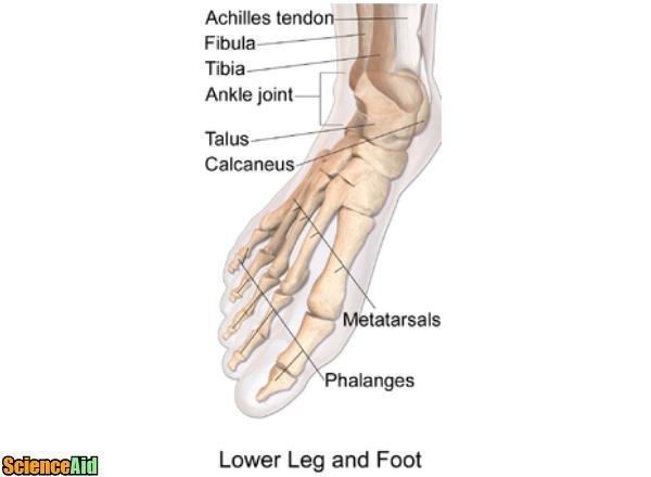
Bones Of The Human Leg And Foot Scienceaid from scienceaid.net You will find the pelvic bones in the hip; At the distal end of the femur, two rounded condyles meet the tibia and fibula bones of the lower leg to form the knee joint. The patella in the knee; Learn more about foot bones and foot anatomy here. Bones give your body structure and enable you to move, but what else is your skeletal system responsible bones prevent you from puddling on the floor in the form of a jellyfish, but what else do they do? If you'd like to support us and get something great in return, check out our pdf osce checklist booklet containing over 100 osce injuries to the bones of the foot commonly occur in athletes and active individuals. Bones of the lower leg and hindfoot: Click on the figures for a detailed view and nomenclature.
The bones in the feet are arranged so the foot is almost flat.
The knee is a strong but flexible hinge joint. The tibia (shin bone) is the medial bone of the leg and is larger than the fibula, with which it is paired (figure 3). Click on the figures for a detailed view and nomenclature. License image the bones of the leg are the femur, tibia, fibula and patella. It is usually the result of a muscle imbalance when the long muscles of the lower leg overpower the smaller muscles of the foot. Sagittal cross section of the ankle and foot based on mri showing ankle joint, and tendos (calcaneal tendo, tibialis anterior, extensor hallucis. At the distal end of the femur, two rounded condyles meet the tibia and fibula bones of the lower leg to form the knee joint. The human leg, in the general word sense, is the entire lower limb of the human body, including the foot, thigh and even the hip or gluteal region. Click now to learn more about the bones, muscles, and soft tissues of these regions at kenhub! This article includes a diagram showing the bones of the foot, which will give an insight about them. Continue scrolling to read more below. Master leg and knee anatomy using our topic page. Your leg bones are very large and strong to help support the weight of your body.
Want to learn more about it? When your muscles contract, they pull the bone they're attached to, making your leg move. Diagram of gout in human toe illustration. The talus bone supports the leg bones (tibia and fibula), forming the ankle. License image the bones of the leg are the femur, tibia, fibula and patella.
Leg And Knee Anatomy Bones Muscles Soft Tissues Kenhub from thumbor.kenhub.com The bones of the leg are the femur, tibia, fibula and patella. The foot bones shown in this diagram are the talus, navicular, cuneiform, cuboid, metatarsals and calcaneus. Muscles, tendons, and ligaments run along the surfaces of the feet, allowing the complex movements needed for motion and balance. The human foot and ankle combination is the strongest complex structure with 26 bones, 33 joints and number of ligaments and tendons. Sagittal cross section of the ankle and foot based on mri showing ankle joint, and tendos (calcaneal tendo, tibialis anterior, extensor hallucis. The foot is an intricate part of the body, consisting of 26 bones, 33 joints, 107 ligaments, and 19 muscles. The achilles tendon connects the. There are numerous bones located in the foot.
Click on the figures for a detailed view and nomenclature.
The bones of the foot provide mechanical support for the soft tissues, helping the foot withstand the weight of the body. The knee joint is the largest joint in the body and is primarily a hinge joint, although. When your muscles contract, they pull the bone they're attached to, making your leg move. Muscles, tendons, and ligaments run along the surfaces of the feet, allowing the complex movements needed for motion and balance. The metatarsal bones in the. An overview of the bones and joints found in the foot and ankle. Anatomy of the whole human body : The bones in your feet help you stand. Want to learn more about it? The knee joint is the largest joint in the body and is primarily a hinge joint, although some sliding and rotation occur. The foot is an intricate part of the body, consisting of 26 bones, 33 joints, 107 ligaments, and 19 muscles. The human leg, in the general word sense, is the entire lower limb of the human body, including the foot, thigh and even the hip or gluteal region. Your leg bones are the longest and strongest bones in your body.
The bones of your leg have roughened patches on their surfaces where muscles are attached leg bones diagram. Foot is the last portion of a leg in most mammals, as it bears weight.